This website is intended for an international audience, excluding the UK, United States, Canada and France
This website is intended for an international audience, excluding the UK, United States, Canada and France
Information about the emotional, physical and social challenges of living with NETs.
Steve

If you’re living with neuroendocrine tumours (NETs) and you’re looking for clinics in your region and patient support services, then you’ve come to the right place. Search through the Get Support resources listed here to see if any of these can help you.

Diagnostic imaging (scans) lets doctors look inside your body for clues about a medical condition. A variety of machines and techniques can create pictures of the structures and activities inside your body.
The main imaging techniques used for diagnosing and monitoring of neuroendocrine tumours (NETs) include ultrasound (sonography), computed tomography (CT), and magnetic resonance imaging (MRI). Positron emission tomography (PET) may also be used.
More specialised imaging techniques include nuclear medicine scans, such as an octreotide scan, Gallium-68 PET scan, bone scintigraphy and an MIBG scan, which are discussed separately.
See the practical tips section for more advice on preparing for imaging scans.
“An abdominal ultrasound or an abdominal computed tomography (CT) scan are useful to evaluate if NETs have spread to the liver”
The healthcare professional shown in this video speaks about their own opinions and experiences and not about any specific patient. Some treatment options may not be authorized or available in your country. Each person’s case is unique and you should always consult a doctor for information and advice about the diagnosis and treatment of NET. No information within this video constitutes medical advice.
“The time between examinations and consultations are stressful. Keep in mind that the disease is very slow in its evolution.”
Ultrasound scans use high frequency sound waves, which people can not hear, to build up a picture of the inside of the body. These scans are completely painless. During an ultrasound test, these sound waves are transmitted through body tissues using an instrument called a transducer. This information is then displayed on a computer monitor.
These scans are usually carried out in the radiology department of a hospital. Two types of ultrasound that may be used to help diagnose NETs include:
CT and MRI are two of the most important imaging techniques for diagnosing NETs. These techniques can both be used to determine the position and size of tumours.
A CT scanner is a special type of X-ray machine that uses radiation to provide a three-dimensional picture of the inside of the body. The CT scan is usually carried out by a radiologist. Regular CT scans are useful to find out more about the rate of tumour growth and how your NETs are responding to treatment.
Before the CT scan, you may be asked to have an injection or drink a fluid containing a ‘contrast agent’ or dye that shows up on the scan. The contrast agent can highlight specific areas inside the body. This can show a clearer image of the results.
During the scan you will need to lie very still for 10 to 20 minutes as the CT scanner passes over you. Unlike an MRI machine the CT scanner does not surround your whole body. It might therefore be more comfortable for people who suffer from a fear of small spaces (claustrophobia).
See the practical tips section for more advice on preparing for imaging scans.
MRI is a type of scan that uses a powerful magnetic field and pulses of radio wave energy to make cross-sectional images of organs, tissues, bones and blood vessels. A computer turns the images into three-dimensional pictures. An MRI scan may be used if an ultrasound scan or CT scan do not provide doctors with enough information to make a diagnosis.
Similar to having a CT scan you will need to lie very still. The scan may take longer (anytime from 30 minutes to up to 1 hour) as you lie inside while the machine scans your body.
A radiologist will then look at these images to determine the difference between normal and diseased tissue. If a tumour is identified in this way, further tests may be needed to confirm the type of NET. Complete results are usually ready for your doctor days or weeks later.

As MRI scans are based on magnetic fields and not X-rays, they are relatively harmless. There are certain preparations and precautions that your doctor should tell you about before the test. For example, you’ll need to remove any metal objects. You will also be given headphones to block out the noise since the scan can be rather loud.
See the practical tips section for more advice on preparing for imaging scans.
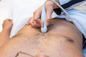
An echocardiography is an imaging test that uses ultrasound to produce moving images of the heart and blood flow through the heart’s valves and structures.
Some types of NETs that are associated with carcinoid syndrome will release hormones called serotonin and tachykinins into the blood stream. These hormones can travel to the heart and affect the cardiac valves.
If you have been diagnosed with carcinoid syndrome your specialist doctor may carry out an echocardiography to examine your heart, and/or a blood test called a NT proBNP, which is used to help detect and evaluate the risk of heart failure.
The echocardiography probe is placed on the chest and images are taken through the chest wall (transthoracic echocardiography).
Depending on the results of the echocardiography, you may need further investigations, such as transesophageal echocardiography or a cardiac MRI.
See the practical tips section for more advice on preparing for imaging scans.
Review the resources used to create the content in this section >
 Diagnosing NETs
This section describes the most common and probable diagnostic or testing methods that you may undergo if your doctor suspects that you have neuroendocrine tumours (NETs).

Learn about the different types of NETs, symptoms of NETs, their diagnosis and treatment.
Learn About NETs
Read about treatment options for NETs, including surgery, radiotherapy and medications.
Treatment Options